
Contents
A qEEG brain map looks impressive: a head outline filled with reds, blues and greens. It is easy to assume the colors hold a clear answer about what is wrong.
The real value is more modest, and more useful once you know its limits. Here is what a qEEG brain mapping session can add to your exam, what it cannot tell you, and how to get a clean recording.
What is a qEEG brain map?
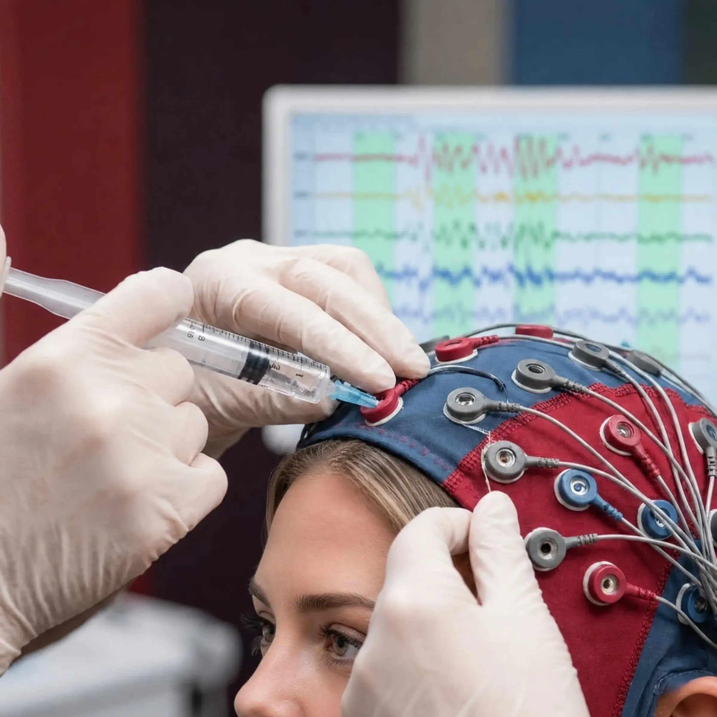
A qEEG, or quantitative EEG, starts with a standard EEG recording of the brain's electrical activity. Software then breaks the signal into frequency bands and turns the numbers into color maps. The maps show how strong each band is at each sensor and how areas relate to each other.
The recording itself is simple. Sensors sit on your scalp with a gel or paste, and they only record. Nothing is sent into your brain, and there is no radiation or enclosed scanner.
You sit still with your eyes open, then closed, while the system records. The analysis happens afterward, on a computer.
What can a brain map show?
A brain map shows patterns in your brainwave activity during the recording: which frequencies are strong or weak, where they appear, and how different areas work together. Those patterns add measured data to your symptoms, history and exam, which on their own are hard to measure.
In practice, a map helps in three ways. It gives a starting point you can compare against later. It points to areas worth a closer look in the exam. It helps choose where brainwave training begins.
- A baseline recorded before any training
- Patterns that line up with, or differ from, your symptoms
- Starting targets for neurofeedback
- A repeat map to compare against, when one is useful
Do red areas on a brain map mean damage?
No. The colors on a brain map show where a value sits higher or lower than the reference the software uses. A red patch is a number that stands out, not a picture of an injury. The exam and your symptoms decide whether that number matters.
This is where many people get worried after seeing their map. A bright area can reflect the moment of the recording, such as tension in the jaw or a late night, as much as anything lasting.
That is why we walk through your map with you, area by area. You see which findings line up with what you feel day to day, and which ones we simply note and recheck later.
What can a brain map not tell you?
A brain map cannot confirm that you have a concussion, ADHD or any other condition. The same pattern can come from different causes, including tiredness, medicines or the way the recording was made. It also cannot show the structure of the brain, which is what an MRI or CT scan does.
Research backs up that caution. A review of 16 studies of athletes after a sports concussion found that every study reported some change in resting brainwaves. The changes differed from study to study, and methods varied too much to set a standard.
A meta-analysis of the theta/beta ratio, a pattern often linked with ADHD, reached a similar view. The ratio was not a reliable way to identify ADHD, although it did stand out in a subgroup of people.
Keep in mind
A physician names a condition; a brain map is one input to that picture, never the whole answer.
The athlete findings come from a 2019 systematic review in the Journal of Neurotrauma.
The theta/beta ratio findings were published as a meta-analysis in the Journal of Attention Disorders.
How is a qEEG different from an MRI or a hospital EEG?
An MRI or CT scan shows the brain's structure, such as bleeding or swelling. A hospital EEG is usually read by eye, often to look for seizure activity. A qEEG uses the same kind of recording but adds math, comparing frequency patterns across the head.
Each test answers a different question. If your physician needs to rule out a bleed or a structural problem, imaging comes first, and a brain map is no substitute.
A qEEG earns its place later, when the question is how your brain activity behaves and how training might change it.
In the U.S., the FDA groups software that shows the frequency content of an EEG as a Class I device, the category covered by general controls. That describes the tool, not a promise about what any map means for you.
The classification is set out in the FDA rule for EEG signal spectrum analyzers, 21 CFR 882.1420.
How do we use a brain map at True Performance?
Your map is one part of a larger evaluation led by Dr. Jeff McWhorter, D.C., a chiropractor whose practice focuses on functional neurology. We read it next to your history, balance and thinking tests, and the goals you bring.
When neurofeedback fits, the map helps set where training starts, as part of our neurofeedback and biofeedback program. If your map and your symptoms tell different stories, we trust the whole picture, not the map alone.
How should you prepare for a qEEG recording?
Wash your hair the night before and skip conditioner and styling products, since they can affect how the sensors work. Tell us about every medicine you take. On the day, try to stay relaxed and still, because blinking and moving can blur the recording.
Sleep matters too. A very short night can change how your brainwaves look, so tell us if you slept badly. If you have a seizure disorder or a scalp wound, mention it on your intro call so we can plan around it.
For more on the recording and how to prepare, see Cleveland Clinic's guide to the EEG.
What should you expect from a brain map?
Expect measured data, not a verdict. A good map adds a baseline, sharpens the questions in your exam and helps plan neurofeedback.
Before a recording, it is fair to ask who will read your map, how it could change your plan, and whether a repeat map will be compared with the first. Clear answers are a good sign.
Be wary of any claim that a map alone can name a condition or promise a result. If you want to see what a map could add to your plan, start with a complimentary 15-minute intro call and read how brain mapping fits into an evaluation here.
Frequently asked questions
Does a qEEG brain map hurt?
No. The sensors rest on your scalp with a gel or paste and only record. Most people find the hardest part is sitting still.
Can a brain map show whether I have ADHD?
No. Research on brainwave patterns linked with ADHD found they are not a reliable way to identify it. A physician makes that call from a full evaluation, and we read the map with your whole exam.
Is a qEEG the same as neurofeedback?
No. A qEEG records and analyzes your brainwaves once. Neurofeedback is a series of training sessions, and the map can help choose where that training starts.
Is a qEEG recording safe for children?
The recording only listens to brain activity, so nothing is sent into the body. We work with patients of all ages, and parents take part in the full evaluation and give written consent.
Does insurance cover qEEG brain mapping?
We explain cost and payment options on your intro call, before you book an exam. What is included depends on your evaluation.
See what a brain map can add to your plan
Start with a complimentary 15-minute call with our team.